Veterinary Necropsy Form PDF Details

For veterinary professionals, having a necropsy form at the ready is essential for any postmortem examination. A properly filled out necropsy report allows veterinarians to document their findings so that they can quickly ascertain causes of death and provide accurate results for clients. However, navigating the different items on the form may seem like an intimidating task at first glance! In this blog post, we'll provide a comprehensive overview of what you should include in your Veterinary Necropsy Form – ultimately enabling you to perform your duties with confidence.

QuestionAnswer
Form NameVeterinary Necropsy Form
Form Length12 pages
Fillable?No
Fillable fields0
Avg. time to fill out3 min
Other namesnecropsy report online, form 1626 form, necropsy report, form 1626

Form Preview Example

VETERINARY NECROPSY REPORT CHECKLIST AND GUIDELINES

(DD Form 1626 may be used independently of TB Med 283. Instructions for completing this form begin on page 9.)

SECTION I - ADMINISTRATIVE DATA

PART A - CONTRIBUTOR'S DATA

1.

CONTRIBUTOR/PROSECTOR

2.

DATE OF REPORT (YYYYMMMDD)


3. NAME AND ADDRESS OF REPORTING UNIT

4.

GEOGRAPHIC LOCATION (Country)

5. TELEPHONE NUMBER

6. FAX NUMBER

7. E-MAIL

PART B - ANIMAL IDENTIFICATION AND RELATED DATA

8. ANIMAL I.D. (Name and Tattoo Number)

9.

SPECIES


10.

BREED


11.

DATE OF BIRTH (YYYYMMMDD)

12. AGE


13. SEX

14. NEUTERED

15.

WEIGHT

16. COLOR


YES


NO


17.

EUTHANIZED (Specify method and agent used.)


YES


NO


18.CAUSE OF DEATH (Medical reason for death or decision to euthanize.)

19.NAME AND ADDRESS OF UNIT ACCOUNTABLE FOR ANIMAL

20. CONTRIBUTOR'S NECROPSY NUMBER

21. DATE OF DEATH (YYYYMMMDD)

22.TIME BETWEEN DEATH AND NECROPSY

23. PRIORITY REQUIRED

ROUTINE


RUSH


24. MATERIALS FORWARDED

SECTION II - CLINICAL AND PATHOLOGICAL DATA

25.CLINICAL ABSTRACT (Continue in Block 32 or on a separate sheet if necessary and attach copy of DD Form 1743, Death Certificate of a Military Dog; DD Form 1834, Military Working Dog Service Record (Assignment History); and DD Form 2619, Master Problem List.)

DD FORM 1626, OCT 2001

PREVIOUS EDITION IS OBSOLETE.

Page 1 of 12 Pages

Adobe Professional 7.0

SECTION II - CLINICAL AND PATHOLOGICAL DATA (Continued)

26.CLINICAL DIAGNOSES (Relevant to the death of the animal.)

27. GROSS NECROPSY DIAGNOSES

28.GROSS PHOTOGRAPHS (Tissues and lesions photographed.)

29.MICROBIOLOGICAL CULTURE RESULTS (Specify site.)

30.CLINICAL PATHOLOGY TEST RESULTS (Relevant to the death of the animal, include a copy.)

31.RADIOGRAPHS AND INTERPRETATIONS (Relevant to the death of the animal, include a copy.)

32.REMARKS (List any additional information that supplements this report. This block can be used for a continuation of another information block.)

33.SIGNATURE OF CONTRIBUTOR/PROSECTOR

DD FORM 1626, OCT 2001

Page 2 of 12 Pages

SECTION III - GROSS FINDINGS

(If more space is needed, identify the tissue and continue on a blank sheet.)

GENERAL (Condition of cadaver, haircoat, body orifices, scars, superficial lesions/tumors, etc.)

WEIGHT

VentralDorsal

EYES AND EARS

PRIMARY INCISION (Subcutaneous fat, musculature, superficial lymph nodes, etc.)

BONE MARROW (Color, consistency, submit two unstained cytology smears.)

ENDOCRINE GLANDS (Pituitary, thyroid, parathyroids, adrenals.)

Thyroid/Parathyroid (gm) L


R

Adrenal (gm) L

R

Pituitary

DD FORM 1626, OCT 2001

Page 3 of 12 Pages

SECTION III - GROSS FINDINGS (Continued)

BODY CAVITIES (If fluid is present in the abdomen and/or thorax, describe the color, clarity and amount.)

URINARY SYSTEM (Kidneys, ureters, urinary bladder, urethra.)

Kidney (gm) L


R

GENITAL SYSTEM (Testes, epididymides, spermatic cords, prostate gland, penis, ovaries, oviducts, uterus, cervix, vagina, vulva.)

ILIAC LYMPH NODES AND ABDOMINAL AORTA

HEART (Pericardium, epicardium, myocardium, endocardium, valves, coronary vessels, etc.) Weight (gm)

RV (mm)

LV (mm)

IV Septum (mm)

VASCULATURE (Arteries, veins, and lymphatics.)

DD FORM 1626, OCT 2001

Page 4 of 12 Pages

SECTION III - GROSS FINDINGS (Continued)

RESPIRATORY SYSTEM (Larynx, trachea, bronchi, lymph nodes, etc.)

LUNG (gm) R

L

LIVER (Size, color, consistency, gallbladder, bile ducts, etc.)

Weight (gm)

PANCREAS

Weight (gm)

SPLEEN

Weight (gm)

GASTROINTESTINAL TRACT (Mouth, teeth, tongue, salivary glands, esophagus, stomach, duodenum, jejunum, ileum, cecum, colon, rectum, anus and lymph nodes.)

DD FORM 1626, OCT 2001

Page 5 of 12 Pages

SECTION III - GROSS FINDINGS (Continued)

BONES AND JOINTS, NONVERTEBRAL (Hip, stifle, shoulder, elbow, other.)

BRAIN (Cerebrum, cerebellum, brainstem.)

NASAL CAVITY AND SINUSES

VERTEBRAL COLUMN

SPINAL CORD

PERIPHERAL NERVES

DD FORM 1626, OCT 2001

Page 6 of 12 Pages

SECTION IV - TISSUE CHECKLIST

Use this checklist to record all tissues collected for submission. In the "Other" section, list any additional tissues collected that are not on the checklist. Tissues are listed in order of descriptive protocol. See TB MED 283 for an alphabetical list.

34. MILITARY WORKING DOG (MWD) NAME

35. MWD TATTOO

36. ITEM


EYES (LEFT/RIGHT)


SPLEEN


LACRIMAL GLAND


MESENTERIC LYMPH NODE(S) *


AXILLARY LYMPH NODE(S) (LEFT/RIGHT) *


STOMACH


HAIRED SKIN (SPECIFY SITE IN REMARKS)


DUODENUM


MAMMARY TISSUE (IF APPLICABLE)


JEJUNUM


SKELETAL MUSCLE


ILEUM


BONE MARROW


ILEOCECOCOLIC JUNCTION


THYROID AND PARATHYROID GLANDS *


CECUM


MANDIBULAR SALIVARY GLANDS


COLON


ADRENAL GLANDS (LEFT/RIGHT)


RECTUM


THYMUS/THYMIC REMNANTS *


ANUS/PERIANAL AREA/ANAL SAC


BONE (RIB, STERNEBRA; SPECIFY OTHER SITE)


EAR CANAL


TONSILS (LEFT/RIGHT) *


BRAIN (INTACT)


TONGUE


PITUITARY GLAND *


ESOPHAGUS


SPINAL CORD (INTACT)


MEDIAL RETROPHARYNGEAL LYMPH NODE(S) *


PERIPHERAL NERVE (SPECIFY)


DIAPHRAGM


OTHER (SPECIFY)


KIDNEYS (LEFT/RIGHT)


URETERS


URINARY BLADDER


URETHRA


PROSTATE GLAND

REMARKS:


TESTES/EPIDIDYMIDES (LEFT/RIGHT)


UTERUS; CERVIX; VAGINA


OVARIES (LEFT/RIGHT)


ILIAC LYMPH NODE(S) (LEFT/RIGHT) *


AORTA, ABDOMINAL (SPECIFY OTHER SITE)


HEART (ENTIRE ORGAN, OPENED)


TRACHEA/LARYNX


TRACHEOBRONCHIAL LYMPH NODE(S) (L/R) *


LUNG


PANCREAS


LIVER


GALLBLADDER


* Identify these tissues by placing in labeled, large-size tissue cassettes (or other containers) prior to fixation and shipment.

DD FORM 1626, OCT 2001

Page 7 of 12 Pages

DISSECTION AIDS

Right Ventricle

Left Ventricle

Aortic Outflow


DISSECTION OF THE HEART


REMOVAL OF THE BRAIN


Cervical

Thoracic

Lumbar

REMOVAL OF THE SPINAL CORD

DD FORM 1626, OCT 2001

Page 8 of 12 Pages

INSTRUCTIONS FOR COMPLETING DD FORM 1626

PURPOSE: DD Form 1626 is used to record necropsy results of military working dogs, but may be used for other animals.

GENERAL: This form may be printed or photocopied without the instruction pages to minimize the length. DD Form 1626 is available on the Army Electronic Library CD-Rom (EM 0001) and on the DoD web site at http://web1.whs.osd.mil/icdhome/icdhome.htm.

SECTION I - ADMINISTRATIVE DATA

1.Blocks 1 through 7: Name and Address of the Reporting Veterinary Unit. Enter the rank, full name and title of the contributing Veterinary Corps officer and the military unit to which assigned. Station address, telephone and fax numbers, and e-mail address are all important information.

2.Blocks 8 through 16: Animal Identification.

3.Block 17: Euthanized (Yes or No). Specify the brand name or the active agent of the euthanasia product, the amount given, and the site of intravenous administration.

4.Block 18: Cause of Death. Concisely state the medical reason(s) leading to the death of the animal. In the case of euthanasia, state why the animal was euthanized.

5.Block 19: Name and Address of Unit Accountable for Animal. Enter the military unit that is accountable for the MWD. Include the complete military unit designation and station address. (If the form is used for a privately owned pet, the owner's information should be indicated in this block.)

6.Block 24: Materials Forwarded. List categories such as tissues, culture results and radiographs, gross photographs, etc.

SECTION II - CLINICAL AND PATHOLOGICAL DATA

7.Block 25: Clinical Abstract. The clinical abstract should address the recent pertinent information regarding this case, and previous significant medical history. Include illnesses, recent treatments, laboratory and radiologic findings, surgeries, wounds, fractures, immunizations, working environment and recent TDY assignments. Attach a copy of DD Form 1743, Death Certificate of a Military Dog; DD Form 1834, Military Working Dog Service Record (Assignment History); and DD Form 2619, Master Problem List.

8.Block 26: Clinical Diagnoses. Concisely list significant clinical conditions relating to the death of this animal.

9.Block 27: Gross Necropsy Diagnoses. List all necropsy diagnoses (interpretations).

10.Block 30: Clinical Pathology Tests and Results. List tests and include a copy of the results.

SECTION III - GROSS FINDINGS

NECROPSY PROTOCOL (Abstract of TB MED 283, Chapter 3)

INTRODUCTION (Para. 3-1)

a.Describe all abnormalities in detail. Use each block/diagram in this section for a systematic examination and report. Enter "NGLR" (no gross lesions recognized) or an equivalent notation as appropriate.

b.Use physiologic saline to rinse tissues during the necropsy. Tissue specimens are sensitive to hydrostatic and osmotic pressures. Rinsing with tap water will cause morphologic artifacts.

c.Organ weight data is optional. Collect when practicable.

d.Use the tissue checklist on Page 7.

EXTERNAL EXAM (Para. 3-2)

Carefully examine all external structures and surfaces and record the following: weight, nutritional condition, state of rigidity, extent of post-mortem decomposition, and the condition of the haircoat, skin, mucous membranes, eyes and body orifices. Describe tattoos, scars, wounds, cutaneous tumors, bony malformations, discharges, etc.

EYES (Para. 3-3)

The eyes should be collected early in the necropsy. Clamp the eyelids together with tissue forceps (Allis recommended). Use a scalpel to incise the eyelids around the orbit. With curved scissors, dissect the eyes from the orbits by cutting extraocular muscles, connective tissue attachments and optic nerve. Examine and collect lacrimal gland tissue. Check for eyelid and retrobulbar lesions. Eyelids should remain attached to the left eye for identification; however, remove skeletal muscle from the surface of the sclera to facilitate fixation. Also, 0.2 - 0.5 ml of formalin may be injected directly into the vitreous chamber.

DD FORM 1626, OCT 2001

Page 9 of 12 Pages

INSTRUCTIONS FOR COMPLETING DD FORM 1626 (Continued)

SECTION III - GROSS FINDINGS (Continued)

VENTRAL MIDLINE INCISION (Para. 3-4)

a.With the carcass in dorsal recumbency, make a ventral midline incision that extends from the mandibular symphysis to the pubis. Dorsally reflect the skin from the abdomen, thorax and cervical region to expose underlying tissues and regional lymph nodes. Examine the amount and color of the subcutaneous adipose tissue.

b.Transect the muscles between the scapula and thorax and the soft tissues surrounding the hip joint to allow the pelvic and thoracic limbs to lay flat and maintain the carcass in a steady position.

c.Locate and examine the brachial plexus in the axillary space. Locate, examine and collect the applicable regional lymph nodes.

d.Carefully examine the subcutis and skeletal muscle. Examine and collect a representative specimen of skeletal muscle.

BONE MARROW (Para. 3-6)

Femur, rib, sternum and vertebra are readily accessible sites for collection of histologic and cytologic red bone marrow specimens. The submitted specimen must contain red marrow. Collect early in the necropsy. (See para. 2-9c)

THYROID AND PARATHYROID GLANDS (Para. 3-7)

Careful removal of the muscles ventral to the trachea will expose the thyroid and parathyroid glands. Examine, and collect the thyroid and parathyroid glands.

SALIVARY GLANDS AND LYMPH NODES (Para. 3-8)

Locate and examine the mandibular, parotid and sublingual salivary glands and mandibular lymph nodes. Collect a specimen of mandibular salivary gland and, if indicated, a mandibular lymph node.

BODY CAVITIES (Para. 3-9)

Methodically examine all viscera in situ and observe for correct anatomic size and position. Record any abnormal characteristics of fluids present in either the abdomen or thorax.

a.Abdomen (Para. 3-9a). To completely expose the abdominal viscera, make a ventral midline incision through the abdominal wall from the xyphoid process to the pubis. From the anterior end of this incision, cut laterally along the posterior margin of the last ribs. From the posterior end of the incision, cut laterally just anterior to the pubis.

b.Adrenal Glands (Para. 3-9a). Locate, examine, and collect both adrenal glands.

c.Thorax (Para. 3-9b). Verify negative intrathoracic pressure by observing and carefully listening for the influx of air resulting from a small stab incision made through the tendinous portion of the diaphragm. Using bone cutters, remove the ventral one-third of the rib cage. Examine the mediastinum, pericardium and diaphragm. Examine the parietal and visceral pleura. Examine the ribs and intercostal muscles. Examine and collect the thymus or thymic remnants, if present, in the cranial mediastinum.

d.Thoracic Viscera Removal. Examine the thoracic viscera in situ, then remove en masse. The resultant "block" of thoracic viscera should include tongue, larynx, trachea, esophagus, heart, aorta and lungs. Place on a large cutting board for further dissection (See para. 3-13).

DIAPHRAGM (Para. 3-9c)

Examine and collect a representative specimen of diaphragm.

PELVIC CANAL (Para. 3-9d)

Locate the obturator foramina on the pelvis. Place the tip of bone cutting shears into one foramen and cut the pubis (caudal) and ischium (cranial). Repeat this on the opposite side. By blunt and sharp dissection, remove the freed section of bone to expose the pelvic canal.

UROGENITAL TRACT (Para. 3-9e)

a.Examine the perineum. Incise the skin and subcutaneous tissues around the external genitalia.

b.Free both kidneys from their attachment sites. Bluntly, dissect the ureters and excise at the urinary bladder. On both kidneys, incise and reflect the renal capsule to the hilus and examine the ureter and renal vasculature. Section the kidneys to the renal pelvis, cutting the left kidney longitudinally and the right kidney transversely (for later identification). Extend the renal pelvic incisions into each ureter. Serially section the kidneys and collect representative specimens from both (no more than 0.5 cm thick) that include the renal papilla. Collect a representative section of ureter.

c.Remove remaining urogenital organs. Record ovariohysterectomy or orchiectomy, as applicable, on the DD Form 1626.

DD FORM 1626, OCT 2001

Page 10 of 12 Pages

INSTRUCTIONS FOR COMPLETING DD FORM 1626 (Continued)

SECTION III - GROSS FINDINGS (Continued)

UROGENITAL TRACT (Continued)

d.Examine the urinary bladder, urethra and prostate gland (as applicable). Open the bladder and urethra along their ventral aspects to the os penis (male) or external urethral meatus (female). Closely examine the urethra and trigone of the urinary bladder. Collect representative specimens of urinary bladder, urethra and prostate gland (as applicable).

e.Examine the male genitalia. Collect, label and submit both testes and epididymides in entirety with a segment of each spermatic cord. For proper fixation bisect the testes.

f.Examine the female genitalia. Use scissors to open the vulva, vestibule, vagina, cervix and uterus to the oviducts. Collect representative specimens of these structures and the ovaries.

DIGESTIVE TRACT REMOVAL (Para. 3-9f)

Examine the anus and rectum. Incise the skin and subcutaneous tissues around the anus and remove the abdominal viscera en masse. Place on a large cutting board for further dissection (See para. 3-17).

ILIAC LYMPH NODES AND ABDOMINAL AORTA (Para. 3-11)

Examine iliac lymph nodes and abdominal aorta. Collect representative specimens.

HEART, GREAT VESSELS AND PERICARDIAL SAC (Para. 3-12)

Incise the pericardial sac. Check for abnormal fluid accumulation. Reflect the pericardium over the base of the heart. Examine the base of the heart for neoplastic disease. Examine the great vessels and atria. Examine the epicardial surface and coronary arteries. Carefully dissect the heart (see fig. 3-1 or page 8 of this form) and examine all endocardial surfaces. Collect and submit the entire heart.

RESPIRATORY SYSTEM (Para. 3-13)

a.Trachea. Examine and open the trachea from the larynx to the level of the primary bronchi. Collect representative specimens.

c. Lungs. Palpate the peripheral and hilar areas of all lung lobes. Isolate and infuse the airways and alveolar spaces of the right cranial lung lobe with formalin (see para. 3-13b). Dissect the remaining lung lobes by opening all major bronchi. Collect representative specimens.

LIVER AND PANCREAS (Para. 3-14)

Examine and collect a representative specimen of pancreas with an attached segment of mesentery and duodenum. Verify the patency of the bile duct by applying pressure to the gallbladder, while observing for bile expulsion at the major duodenal papilla. Open and examine the gallbladder. Open the large hepatic arteries and veins (visceral surface) and examine. Serially section the liver and collect representative specimens (no more than 0.5 cm thick) of liver and gallbladder.

SPLEEN (Para. 3-15)

Examine, serially section and collect one or more representative specimens (no more than 0.5 cm thick).

MESENTERY (Para. 3-16)

Examine the omentum, mesentery and the root of the mesentery. Examine and collect representative mesenteric lymph nodes.

GASTROINTESTINAL TRACT (Para. 3-17)

a.Oral Cavity. Examine the oral cavity, teeth, pharynx and larynx. Locate, examine and collect each tonsil. Sharp dissection through the soft tissue between the tongue and mandible, across the soft palate and through the hyoid apparatus will free the tongue and oropharynx. Locate, examine and collect the medial retropharyngeal lymph nodes.

b.Salivary Glands and Lymph Nodes. Locate and examine the mandibular, parotid and sublingual salivary glands, and regional lymph nodes. Collect representative specimens of mandibular salivary glands and regional lymph nodes, if indicated. Separately label tissues that have a similar appearance, for later identification.

c.Esophagus (Para. 3-9c). Open, examine and collect a representative specimen of esophagus. The distal esophagus may be ligated with suitable material prior to transection to prevent spillage of gastric contents.

d.Stomach and Intestines. Open, examine and rinse the mucosal surfaces of the distal esophagus, stomach and duodenum with physiologic saline. Do not rinse with tap water. Collect designated specimens. Open, examine and collect representative specimens from the remaining small and large intestine. Alternately, specimens of intestine may be collected by resecting unopened (8-10 cm) segments, gently flushing the lumen with physiologic saline, ligating each end, and then gently distending the segment with formalin using a syringe and needle.

DD FORM 1626, OCT 2001

Page 11 of 12 Pages

INSTRUCTIONS FOR COMPLETING DD FORM 1626 (Continued)

SECTION III - GROSS FINDINGS (Continued)

BONES AND JOINTS, NONVERTEBRAL (Para. 3-5 and 3-18)

Carefully open the hip, stifle, shoulder and elbow joints (and others as indicated) and examine for abnormal synovial fluid, ruptured, stretched or frayed ligaments, erosion and ulceration of articular cartilage, thickened joint capsules, osteophyte formation and proliferative or thickened synovium. Describe all abnormalities. Collect representative specimens for histopathologic examination, as indicated.

BRAIN (Para. 3-19)

a.Remove the head.

b.Make a dorsal midline incision from the nose to the foramen magnum.

c.Reflect the skin ventrally. Transect and collect a specimen of the ear canal. Remove the temporal muscles from the cranium.

d.Use a bone saw to make three cuts through the skull. Do not cut into the brain. The first cut is made transversely at the anterior limit of the cranial cavity, slightly posterior to the zygomatic process. Rotate the cranium to one side and connect the end of the transverse cut with the foramen magnum. Repeat on the opposite side (See figure 3-2 or page 8 of this form).

e.Use a Virchow's skull breaker, bone chisel or similar instrument in the first (transverse) incision, to pry off the calvarium. Examine the internal surface of the calvarium.

f.If necessary, remove the osseous tentorium cerebelli with rongeurs. Transect the olfactory lobes. Elevate the rostrum and carefully transect the cranial nerves and pituitary stalk, freeing the brain.

g.Immerse the brain in formalin (see para. 2-10). Bisect the brain only if tissue is required for immediate laboratory testing, such as for rabies.

PITUITARY GLAND (Para. 3-20)

Incise the dura mater surrounding the sella turcica to free the pituitary gland. Remove it carefully and place in a labeled tissue cassette.

NASAL CAVITY AND SINUSES (Para. 3-21)

Make a complete transverse cut across the frontal and maxillary bones rostral to the orbits. Examine the exposed nasal cavity and sinuses. Alternatively, a wedge of bone may be removed to expose these spaces. If indicated, submit a representative specimen.

VERTEBRAL COLUMN (Para. 3-10)

Examine the ventral surface of the vertebral column and record abnormalities.

SPINAL CORD (Para. 3-22, and Fig. 3-3 or page 8 of this form)

a.Remove the skin remaining on the carcass and examine dorsal subcutis and musculature.

b.Remove the epaxial muscles.

c.Removing the thoracic vertebral arches first allows visualization of the correct placement of the saw blade for the subsequent removal of cervical and lumbar arches. Do not cut into the spinal cord.

d.Thoracic vertebrae. Transect the spinous processes of the thoracic vertebrae with a bone saw. Cut through the vertebral arches adjacent to the remnants of the spinous processes at approximately a 45-degree angle. Make a transverse cut anterior to T-1 and posterior to T-13. Remove the thoracic arches to expose the spinal cord.

e.Lumbar vertebrae. Cut through the vertebral arches immediately dorsal to the transverse processes at a

90-degree angle from vertical (perpendicular to the spinous processes). Make a transverse cut at the lumbosacral junction. Remove the lumbar arches to expose the spinal cord.

f.Cervical vertebrae. Cut through the vertebral arches midway between the spinous processes and the transverse processes at a 0-degree angle (parallel to the spinous processes). Remove the cervical arches to expose the spinal cord.

g.Sacral vertebrae. Opening the sacrum is usually not necessary. If lesions are suspected in the cauda equina, cutting between the intermediate and lateral sacral crests can expose the sacral vertebral canal.

h.Grasp the dura mater with tissue forceps, cut the nerve roots and remove the spinal cord from the canal. Examine the dura mater and spinal nerve roots.

i.To facilitate examination and fixation, carefully cut and reflect the dura mater along the dorsal midline for the full length of the spinal cord.

j.Examine the vertebral column for herniated disc material, osteophyte formation and other lesions.

k.Place a suture through the dura mater (or use some other form of identification) to mark suspect areas of the spinal cord requiring the attention of the histopathologist.

l.Immerse the spinal cord and the attached dura mater in formalin.

PERIPHERAL NERVES (Para. 3-23). Examine and collect radial and sciatic nerve. Staple both ends of the collected specimens to a section of tongue depressor labeled with the tissue identification.

DD FORM 1626, OCT 2001

Page 12 of 12 Pages

How to Edit Veterinary Necropsy Form Online for Free

Working with PDF documents online is actually very easy using our PDF tool. Anyone can fill out veterinary necropsy form here and use several other functions available. To have our tool on the leading edge of convenience, we strive to integrate user-oriented capabilities and improvements on a regular basis. We are always thankful for any suggestions - assist us with remolding how we work with PDF docs. Here is what you would want to do to get going:

Step 1: Press the "Get Form" button in the top part of this webpage to open our PDF tool.

Step 2: When you access the editor, you will see the document ready to be filled in. Apart from filling out various fields, you could also do various other things with the PDF, particularly putting on your own words, changing the initial textual content, adding images, signing the PDF, and much more.

To be able to complete this PDF document, make sure that you enter the required details in each and every area:

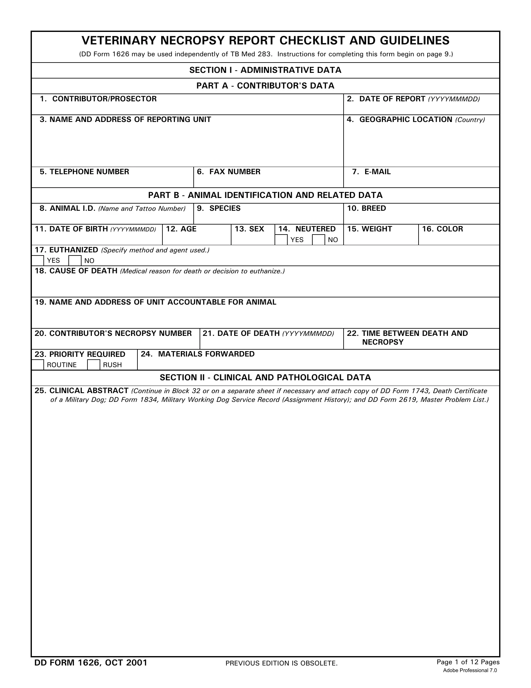
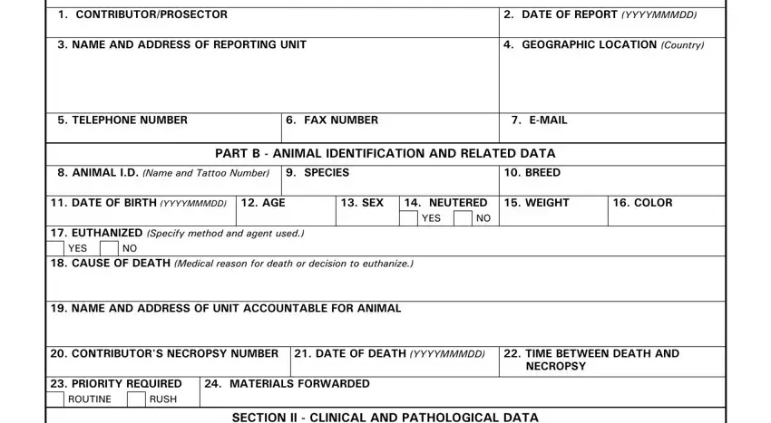
1. The veterinary necropsy form requires certain information to be typed in. Be sure the next fields are complete:

Filling in part 1 of sample of necropsy report

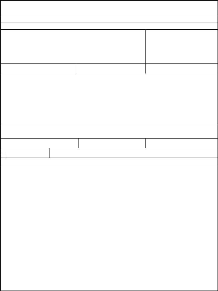
2. The subsequent step is to complete all of the following blank fields: CLINICAL ABSTRACT Continue in.

Find out how to complete sample of necropsy report step 2

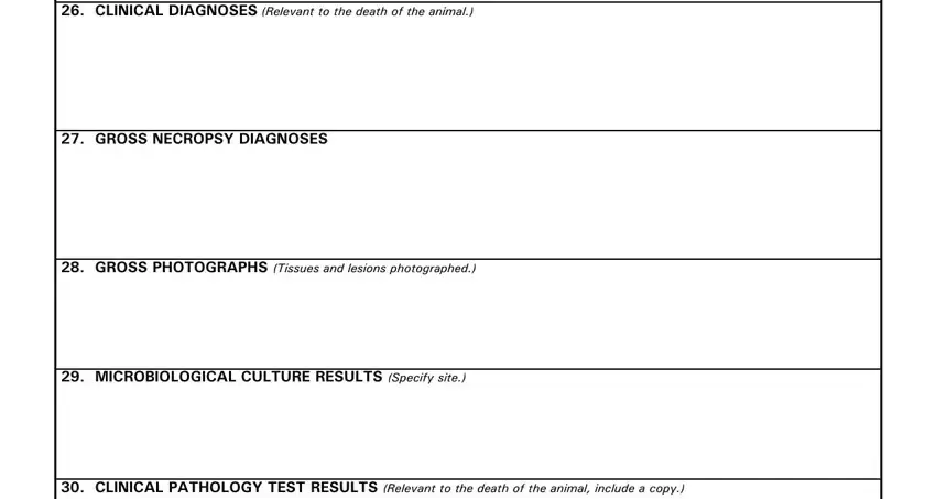
3. Completing CLINICAL DIAGNOSES Relevant to, GROSS NECROPSY DIAGNOSES, GROSS PHOTOGRAPHS Tissues and, MICROBIOLOGICAL CULTURE RESULTS, and CLINICAL PATHOLOGY TEST RESULTS is essential for the next step, make sure to fill them out in their entirety. Don't miss any details!

Filling in segment 3 in sample of necropsy report

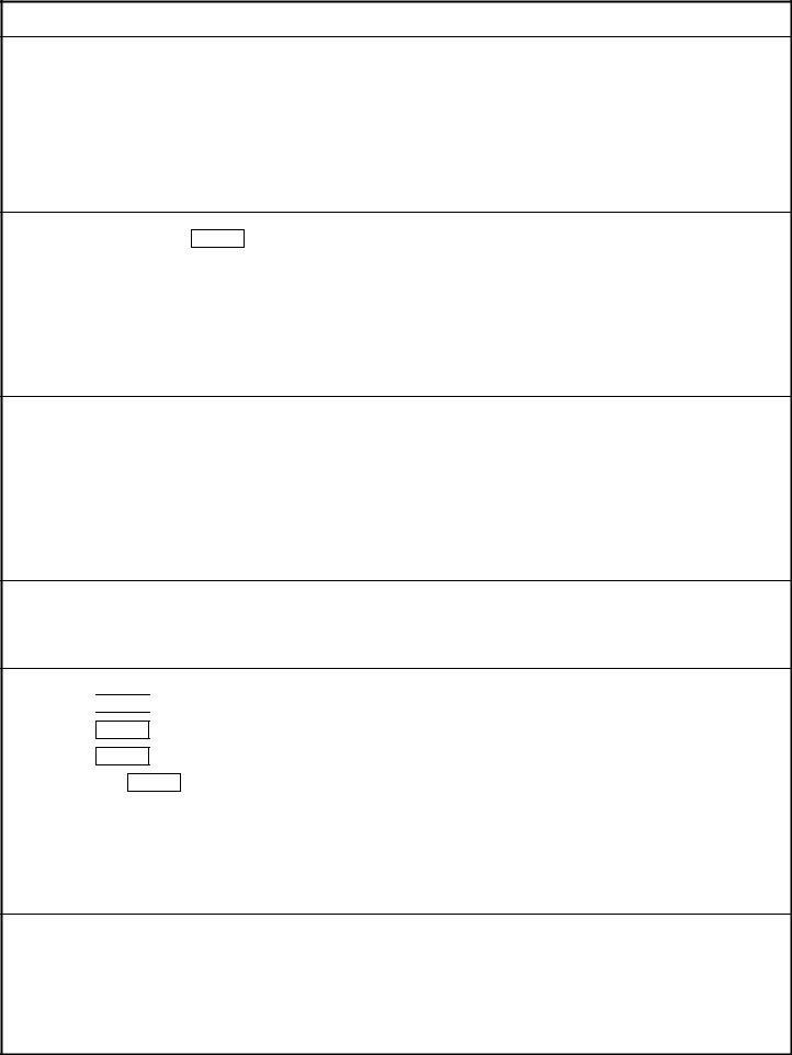
4. This next section requires some additional information. Ensure you complete all the necessary fields - RADIOGRAPHS AND INTERPRETATIONS, REMARKS List any additional, and SIGNATURE OF CONTRIBUTORPROSECTOR - to proceed further in your process!

sample of necropsy report completion process shown (stage 4)

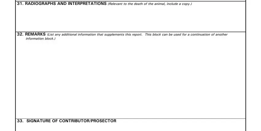
Always be extremely careful when filling out SIGNATURE OF CONTRIBUTORPROSECTOR and REMARKS List any additional, as this is the part where many people make mistakes.

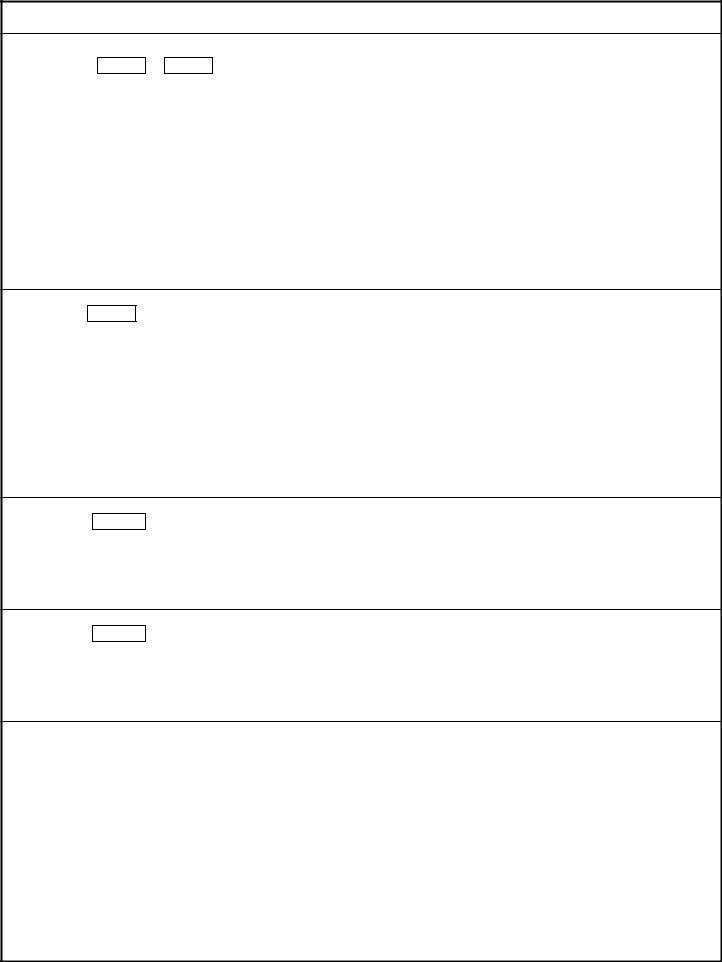
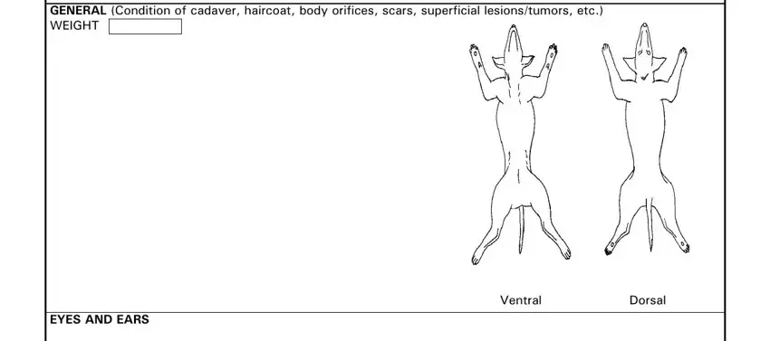
5. Since you approach the finalization of the file, there are a few extra points to complete. Specifically, If more space is needed identify, GENERAL Condition of cadaver, EYES AND EARS, Ventral, and Dorsal must be filled in.

How to prepare sample of necropsy report portion 5

Step 3: Spell-check what you've typed into the blank fields and press the "Done" button. After creating afree trial account at FormsPal, it will be possible to download veterinary necropsy form or email it right away. The PDF will also be at your disposal from your personal cabinet with your edits. FormsPal is devoted to the confidentiality of our users; we make certain that all information used in our system is kept protected.