Exploring the complexities of the human body, especially how it processes the food and drink that sustain us, is an essential aspect of understanding overall health. The HASPI Medical Anatomy & Physiology Lab Activity on the digestive system dives into this intricate process, detailing how digestion breaks down consumables into small, transportable molecules for cellular use. As food traverses the gastrointestinal (GI) tract—a one-way path including the mouth, esophagus, stomach, intestines, and more—it undergoes digestion, absorption, and elimination, with each organ playing a specific role. Specialized tissues and muscles within these organs facilitate movement and chemical breakdown, while the liver, pancreas, and gallbladder contribute vital enzymes and bile. This Lab Activity encompasses not only the journey of digestion but also addresses common digestive disorders, their prevalence and impact on healthcare, and diagnostic methods for identifying these conditions. Through station-based exercises, participants engage with the anatomy and functionality of the digestive system, tracing the GI tract's length and learning about the structure and function of key digestive organs. This comprehensive exploration provides a hands-on approach to understanding one of the body's most fundamental systems.
| Question | Answer |
|---|---|
| Form Name | Haspi Medical Anatomy Physiology Lab Form |
| Form Length | 10 pages |
| Fillable? | No |
| Fillable fields | 0 |
| Avg. time to fill out | 2 min 30 sec |
| Other names | haspi medical anatomy and physiology, haspi digestive system lab answers, digestive system haspi medical anatomy physiology, haspi medical anatomy and physiology answers |
HASPI Medical Anatomy & Physiology 15a
Lab Activity
Name(s): ________________________
Period: _________ Date: ___________
The Digestive System
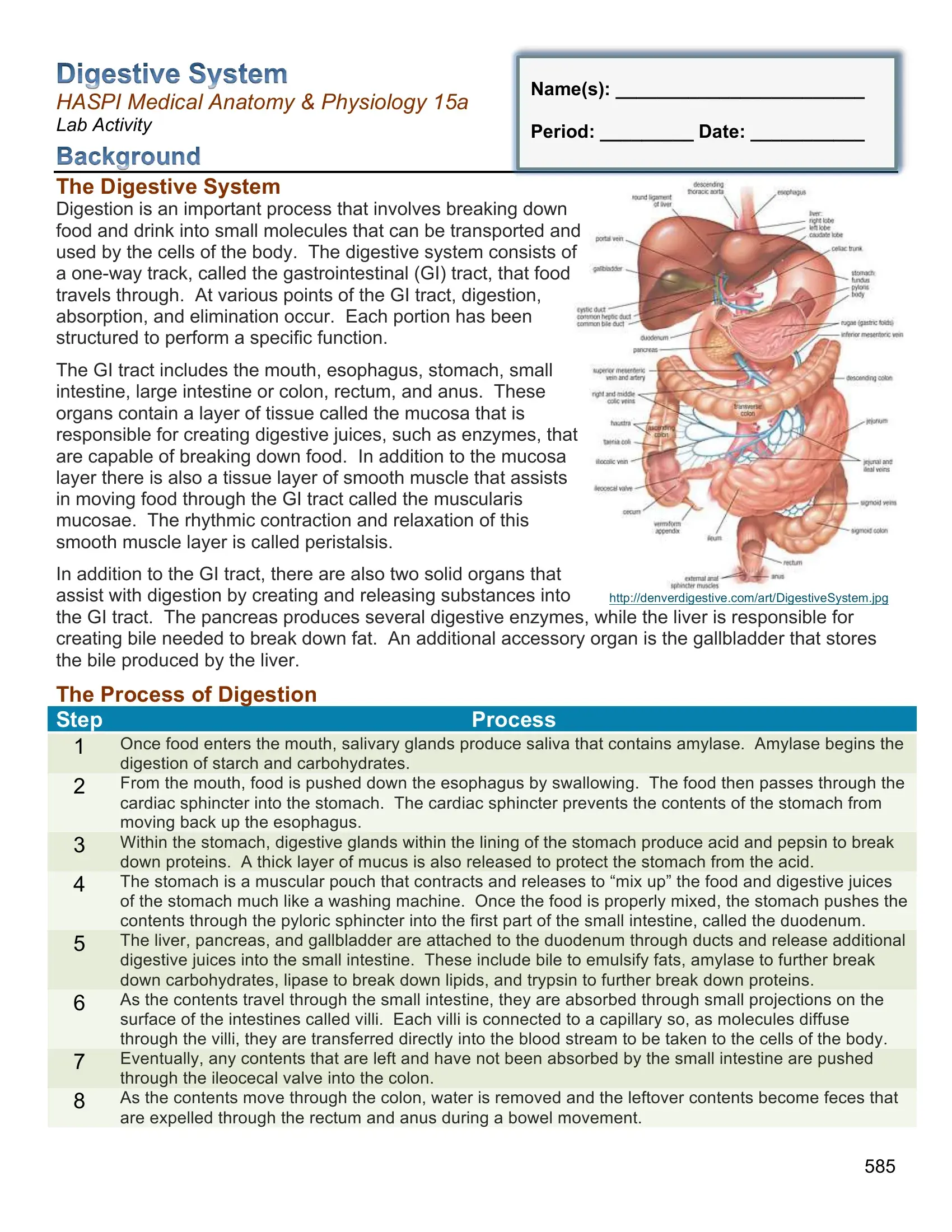
Digestion is an important process that involves breaking down food and drink into small molecules that can be transported and used by the cells of the body. The digestive system consists of a
The GI tract includes the mouth, esophagus, stomach, small intestine, large intestine or colon, rectum, and anus. These organs contain a layer of tissue called the mucosa that is responsible for creating digestive juices, such as enzymes, that are capable of breaking down food. In addition to the mucosa layer there is also a tissue layer of smooth muscle that assists in moving food through the GI tract called the muscularis mucosae. The rhythmic contraction and relaxation of this smooth muscle layer is called peristalsis.
In addition to the GI tract, there are also two solid organs that
assist with digestion by creating and releasing substances into http://denverdigestive.com/art/DigestiveSystem.jpg the GI tract. The pancreas produces several digestive enzymes, while the liver is responsible for creating bile needed to break down fat. An additional accessory organ is the gallbladder that stores the bile produced by the liver.
The Process of Digestion
Step |
Process |
1Once food enters the mouth, salivary glands produce saliva that contains amylase. Amylase begins the digestion of starch and carbohydrates.
2From the mouth, food is pushed down the esophagus by swallowing. The food then passes through the
cardiac sphincter into the stomach. The cardiac sphincter prevents the contents of the stomach from moving back up the esophagus.
3Within the stomach, digestive glands within the lining of the stomach produce acid and pepsin to break
down proteins. A thick layer of mucus is also released to protect the stomach from the acid.
4The stomach is a muscular pouch that contracts and releases to “mix up” the food and digestive juices of the stomach much like a washing machine. Once the food is properly mixed, the stomach pushes the
contents through the pyloric sphincter into the first part of the small intestine, called the duodenum.
5The liver, pancreas, and gallbladder are attached to the duodenum through ducts and release additional digestive juices into the small intestine. These include bile to emulsify fats, amylase to further break down carbohydrates, lipase to break down lipids, and trypsin to further break down proteins.
6As the contents travel through the small intestine, they are absorbed through small projections on the surface of the intestines called villi. Each villi is connected to a capillary so, as molecules diffuse through the villi, they are transferred directly into the blood stream to be taken to the cells of the body.
7Eventually, any contents that are left and have not been absorbed by the small intestine are pushed through the ileocecal valve into the colon.
8As the contents move through the colon, water is removed and the leftover contents become feces that are expelled through the rectum and anus during a bowel movement.
585
Digestive Disorders
There are many diseases and disorders associated with the digestive system. Some of the most important aspects of digestive disorders are the prevalence, healthcare costs, and mortality. Digestive disorders cost the U.S. approximately $141.8 billion dollars in 2004. The following chart summarizes the impact of the most common digestive disorders in the United States in 2004.
|
Digestive |
|
|
Description |
|
|
Diagnosis |
|
|
Healthcare & |
|
|
Annual |
|
|
|
|
|
|
|
|
|
|
|
|||||
|
Disorder |
|
|
|
|
|
|
Hospital Visits |
|
|
Mortality |
|
||
|
|
|
|
|
|
Prevalence |
|
|
|
|
|
|||
|
|
|
|
|
|
|
|
|
|
|
|
Rate |
|
|
|
|
|
|
|
|
|
|
|
|
|
|
|
|
|
|
Constipation |
|
|
Infrequent bowel movements |
|
|
63 million |
|
|
6.9 million |
|
|
137 |
|
|
Diverticulitis |
|
Inflammation of small pouches of the |
|
3.2 million |
|
2.2 million |
3,372 |
|
|||||
|
|
|
|
intestines |
|
|
|
|
|
|
|
|
|
|
|
Gallstones |
|
|
Deposits that form in the gallbladder |
|
|
20 million |
|
|
2.4 million |
|
|
1,092 |
|
|
Gastroesophageal |
|
Stomach contents leak back up |
|
60 million |
|
21.4 million |
1,150 |
|
|||||
|
Reflux (GERD) |
|
through the esophagus |
|
|
|
|
|
|
|
|
|
||
|
|
|
|
|
|
|
|
|
|
|
|
|
||
|
GI Infections |
|
|
May be bacterial, viral, or fungal |
|
|
211 million |
|
|
2.7 million |
|
|
4,396 |
|
|
Hemorrhoids |
|
Swollen veins in the rectum or anus |
75% 45+ |
|
|
3.5 million |
14 |
|
|||||
|
Abdominal |
|
|
Intestines protrude through the |
|
|
4.7 million |
|
|
3.7 million |
|
|
1,663 |
|
|
Hernia |
|
|
abdominal wall |
|
|
|
|
|
|
|
|
|
|
|
Inflammatory Bowel |
|
Inflammation of the intestinal lining |
359,000 |
|
|
1.2 million |
622 |
|
|||||
|
Disease (IBD) |
|
|
|
|
|
|
|
|
|
|
|
|
|
|
Irritable Bowel |
|
|
Irritation of the intestinal lining |
|
|
15.3 million |
|
|
3.2 million |
|
|
20 |
|
|
Syndrome (IBS) |
|
|
|
|
|
|
|
|
|
|
|
|
|
|
Liver Disease |
|
Liver inflammation and tissue damage |
|
2.6 million |
|
3.1 million |
36,090 |
|
|||||
|
Pancreatitis |
|
|
Inflammation of the pancreas |
|
|
1.1 million |
|
|
454,000 |
|
|
3,480 |
|
|
Peptic Ulcers |
|
Sores in the lining of the stomach |
|
14.5 million |
|
1.9 million |
3,000 |
|
|||||
|
Colitis |
|
|
Sores and inflammation of the colon |
|
|
619,000 |
|
|
798,000 |
|
|
311 |
|
|
Viral Hepatitis |
|
Inflammation of the liver caused by the |
|
5.4 million |
|
3.9 million |
5,298 |
|
|||||
|
|
|
|
hepatitis virus |
|
|
|
|
|
|
|
|
|
|
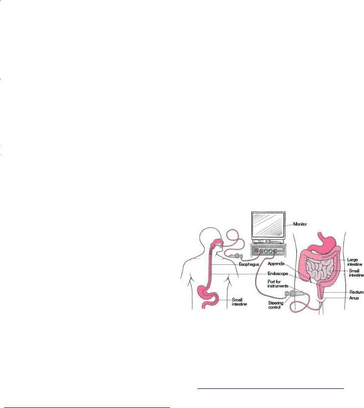
Diagnostic Tests for Digestive Disorders There are many diagnostic tests for digestive disorders. Biopsy samples, gastric juice samples, or stool samples can be used to recognize whether there may be a digestive source for a patient’s symptoms. Following lab tests that may signify a digestive disorder, imaging of the GI tract can be used. The
most effective diagnostic procedures involve using an endoscope to view portions of the GI tract with a camera. When this process is done through the rectum to view the colon, it is called a colonoscopy. When this process is done through the mouth to
view the esophagus, stomach, and/or duodenum, it http://www.merckmanuals.com/media/home/figures/GI_d
is called an EGD or an esophagogastroduodenoscopy. |
igestive_tract_endoscope.gif |
|
Upper and lower GI series can also create radiographic images of the GI tract using a contrast dye, such as barium. The images can show blockages, strictures, or other issues that may exist with the passage of food through the intestines. Using these imaging techniques, healthcare specialists are able to view and identify inflammation, ulcers, growths, and bleeding.
NIH. 2008. Your Digestive System and How it Works. National Institutes of Health (NIH), National Digestive Diseases Information Clearinghouse (NDDIC), NIH Publication No.
NIH. 2010. Digestive Diseases Statistics for the United States. No.
http://digestive.niddk.nih.gov/statistics/statistics.aspx#ack
586