Getting to grips with the Musculoskeletal Examination Ppt form provides a comprehensive overview for anyone keen on mastering the art of musculoskeletal system examination. This guide, authored by Robert Sallis, MD, a notable figure with deep roots in family medicine and sports medicine, offers a treasure trove of insight aimed at both seasoned practitioners and students. Housed within the pages are detailed evaluations of various body parts including the shoulder, neck, elbow, wrist, hand, back, hip, knee, and ankle. Each section is meticulously designed to enhance understanding of examination skills, highlighting crucial landmarks and essential maneuvers to pinpoint potential issues effectively. As an educational tool supplemented by the American Academy of Family Physicians, the material is not only a testament to Dr. Sallis’s expertise but also reflects a collective knowledge shared by contributing experts in the field. The form further encourages the pursuit of formal courses and clinical experience to solidify these examination capabilities. For medical professionals aiming to refine their diagnostic skills, engaging with this form is a step toward achieving excellence in patient care.
| Question | Answer |
|---|---|
| Form Name | Musculoskeletal Examination Ppt Form |
| Form Length | 87 pages |
| Fillable? | No |
| Fillable fields | 0 |
| Avg. time to fill out | 21 min 45 sec |
| Other names | examination skills musculoskeletal, musculoskeletal assessment form, physicians examination musculoskeletal, physicians examination musculoskeletal make |
Examination
Skills of the
Musculoskeletal
System
Author:
Robert Sallis, MD, FAAFP, FACSM
Department of Family Medicine
Kaiser Permanente Medical Center
Fontana, California
Examination Skills of the Musculoskeletal System
Table of Contents
Disclaimer . . . . . . . . . . . . . . . . . . . . . . . . . . . . . . . . . . . . . . . . . . 3
Author Disclosure . . . . . . . . . . . . . . . . . . . . . . . . . . . . . . . . . . . . 3
About the Author . . . . . . . . . . . . . . . . . . . . . . . . . . . . . . . . . . . . . 3
Contributors. . . . . . . . . . . . . . . . . . . . . . . . . . . . . . . . . . . . . . . . . 4
CME Credit Information . . . . . . . . . . . . . . . . . . . . . . . . . . . . . . . . 4
Objectives . . . . . . . . . . . . . . . . . . . . . . . . . . . . . . . . . . . . . . . . . . 5
Introduction . . . . . . . . . . . . . . . . . . . . . . . . . . . . . . . . . . . . . . . . . 6
Shoulder and Neck Evaluation . . . . . . . . . . . . . . . . . . . . . . . . . . . 7
Shoulder and Neck Exam Landmarks . . . . . . . . . . . . . . . . . . 16
Shoulder and Neck Exam Essentials . . . . . . . . . . . . . . . . . . 17
Elbow Evaluation . . . . . . . . . . . . . . . . . . . . . . . . . . . . . . . . . . . . 19
Elbow Exam Landmarks . . . . . . . . . . . . . . . . . . . . . . . . . . . . 26
Elbow Exam Essentials . . . . . . . . . . . . . . . . . . . . . . . . . . . . 27
Wrist and Hand Evaluation. . . . . . . . . . . . . . . . . . . . . . . . . . . . . 29
Wrist and Hand Exam Landmarks. . . . . . . . . . . . . . . . . . . . . 37
Wrist and Hand Exam Essentials . . . . . . . . . . . . . . . . . . . . . 38
Lower Back Evaluation . . . . . . . . . . . . . . . . . . . . . . . . . . . . . . . 39
Lower Back Exam Landmarks . . . . . . . . . . . . . . . . . . . . . . . 45
Lower Back Exam Essentials . . . . . . . . . . . . . . . . . . . . . . . . 46
Hip Evaluation . . . . . . . . . . . . . . . . . . . . . . . . . . . . . . . . . . . . . . 47
Hip Exam Landmarks . . . . . . . . . . . . . . . . . . . . . . . . . . . . . . 54
Hip Exam Essentials. . . . . . . . . . . . . . . . . . . . . . . . . . . . . . . 55
Knee Evaluation . . . . . . . . . . . . . . . . . . . . . . . . . . . . . . . . . . . . 57
Knee Exam Landmarks . . . . . . . . . . . . . . . . . . . . . . . . . . . . 66
Knee Exam Essentials . . . . . . . . . . . . . . . . . . . . . . . . . . . . . 67
Ankle Evaluation . . . . . . . . . . . . . . . . . . . . . . . . . . . . . . . . . . . . 69
Ankle Exam Landmarks . . . . . . . . . . . . . . . . . . . . . . . . . . . . 76
Ankle Exam Essentials . . . . . . . . . . . . . . . . . . . . . . . . . . . . . 77
References . . . . . . . . . . . . . . . . . . . . . . . . . . . . . . . . . . . . . . . . 79
Acknowledgements . . . . . . . . . . . . . . . . . . . . . . . . . . . . . . . . . . 79
Ð 2 Ð
Examination Skills of the Musculoskeletal System
DISCLAIMER
The material presented in this program is being made available by the American Academy
of Family Physicians for educational purposes only. This material is not intended to represent the only, nor necessarily best, methods or procedures appropriate for the medical situations discussed, but rather is intended to present an approach, view, statement or opinion of the faculty that may be helpful to others who face similar situations.
Physicians may choose to check specific details such as drug doses and contraindications, etc., in standard sources prior to clinical application. Every effort has been made to assure the accuracy of the data presented in this program.
This program should be used as an educational tool to help learn the procedures involved in the examination of the musculoskeletal system. In addition to this program, attendance at a for- mal course related to these skills and supervised clinical experience is highly recommended.
AUTHOR DISCLOSURE
The AAFP has selected and provides funding for all authors of this syllabus. According to AAFP policy, all relationships between speakers and proprietary entities will be disclosed.
The following author has returned a disclosure form indicating the following:
Robert Sallis, M.D. Ð Member, Sports Medicine Review Board,
Gatorade Sports Science Institute
The above author has declared that the content of his presentation will not include discussion of unapproved or investigational uses of products or devices.
ABOUT THE AUTHOR
Robert Sallis, MD, FAAFP, FACSM serves as
Ð 3 Ð
Examination Skills of the Musculoskeletal System
CONTRIBUTORS
Author:
Robert E. Sallis, M.D., FAAFP,
AAFP Project Manager:
Sandy Shelton, CME Production Manager, Division of Continuing Medical Education
AAFP Staff Editor:
Cara Sloan, Senior CME Projects Editor, Division of Continuing Medical Education
AAFP CME Projects Coordinator:
Nina Carnoali, Senior CME Program Coordinator, Division of Continuing Medical Education
AAFP Editorial Assistant:
Julie McNamara, Manager, CME Marketing, Division of Continuing Medical Education
Illustrator:
Stephen Beebe, Kaiser Permanente, Los Angeles, California
Videographer:
Rick Tilley, Kaiser Permanente, Los Angeles, California
CME CREDIT INFORMATION
See insert for CME credit information and credit statement.
AAFP members wishing to obtain CME credit should complete and return the enclosed post- test to the CME Production Department, American Academy of Family Physicians, 11400 Tomahawk Creek Parkway, Leawood, Kansas
Physicians who are nonmembers should complete the
Ð 4 Ð
Examination Skills of the Musculoskeletal System
OBJECTIVES
Upon completion of this program, you should be able to:
1.State important history questions used to evaluate patients presenting with problems involving the shoulder, neck, elbow, wrist, hand, back, knee, foot and ankle.
2.Locate important anatomic landmarks in each of these areas and understand their clinical significance.
3.Perform essential exam maneuvers needed to effectively diagnose problems involving the shoulder, neck, elbow, wrist, hand, back, knee, foot and ankle.
Ð 5 Ð
Examination Skills of the Musculoskeletal System
INTRODUCTION
This video presentation is designed to give primary care physicians a practical review of essential examination techniques necessary to diagnose patients presenting with common musculoskeletal complaints. A systematic approach is presented, which includes a focused history followed by a thorough physical exam. The techniques reviewed in this presentation should enable the primary care physician to make confident evaluations and diagnoses.
This
1.Neck/shoulder
2.Elbow
3.Wrist/hand
4.Lower back
5.Hip
6.Knee
7.Ankle
To maximize learning, it is advised you watch the video presentation with a partner while carefully reviewing this syllabus material. At the end of each module, locate each of the anatomic landmarks on your partner (it may be helpful to mark them with an erasable pen). Next, review the list of Òexam essentialsÓ for each module and practice them on your partner.
You may consider viewing the companion ÒJoint Injection and AspirationÓ
Terminology
Before you begin this module, it may be helpful to review terms that will be used to describe various exam maneuvers and findings:
1.Valgus Ñ describes the position of a joint when the distal segment is angled away from the midline of the body (eg, genu valgum is the
2.Varus Ñ the opposite of valgus, in which the distal segment is angled toward the midline of the body (eg, genu varum is the
3.Abduction Ñ refers to motion away from the midline of the body.
4.Adduction Ñ refers to motion toward the midline of the body.
5.Proximal Ñ means closer to any point of reference; opposed to distal.
6.Distal Ñ means further from any point of reference; opposed to proximal.
7.Volar Ñ the palmar surface of the hand.
8.Dorsal Ñ the posterior surface (or back) of the hand.
Ð 6 Ð

Examination Skills of the Musculoskeletal System
Shoulder and Neck
Evaluation
Shoulder and Neck Exam Landmarks
Shoulder and Neck Exam Essentials
Ð 7 Ð
Examination Skills of the Musculoskeletal System
Ð 8 Ð
Examination Skills of the Musculoskeletal System
SHOULDER AND NECK EVALUATION
HISTORY
A thorough history is critical in evaluating patients with shoulder pain. Important questions include:
What was the mechanism of injury or overuse?
It is important to determine if this is a chronic injury related to overuse, or an acute injury related to trauma. Specifically ask what activities cause the pain. Most commonly, pain from an overuse injury will be related to repetitive overhead activity and will tend to worsen with activity and improve with rest. Keep in mind also that pain in the shoulder can radiate from a variety of sources, including the chest, abdomen and the cervical spine.
Are there symptoms of instability?
Ask the patient if they have ever had a dislocated shoulder. This injury will generally result in loosening of the static restraints of the shoulder (capsule and glenohumeral ligaments) and chronic problems of shoulder instability. Inquire if the shoulder Òslips out of placeÓ with throwing or other overhead motions. This is an obvious sign of glenohumeral instability. Instability is commonly seen in young, active patients with recurrent shoulder pain.
What is the location and character of pain?
Asking about the location of pain can be helpful in pinpointing its source, and can be confirmed by palpation. The character of the pain can be helpful in diagnosing rotator cuff problems. With rotator cuff tendinitis, the pain tends to worsen with activity, improve with rest and is typically located in the subacromial area. Pain from impingement syndrome is worse with overhead motions (such as washing hair or reaching for an overhead cupboard). Patients will often wake at night when rolling over onto an extended arm. Finally, pain from a rotator cuff tear will pres- ent as a dull, unrelenting ache
Are there mechanical symptoms (locking or popping)?
Popping or snapping in the shoulder with overhead motion is common but rarely of clinical significance. However, when it is painful or leads to a true blocking of motion, a labrum tear should be suspected.
What is the relationship of pain to the throwing motion?
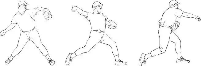
Repetitive throwing commonly causes shoulder pain. The throwing motion can be simply divid- ed into three phases: (1) cocking, (2) acceleration and (3) release/deceleration (Figure 1). Where in the throwing motion the pain occurs can be a clue to its cause. Pain during the cock- ing phase suggests anterior cuff tendinitis or anterior instability/subluxation. Pain during the acceleration phase suggests rotator cuff tendinitis or impingement. Pain during release/decelera- tion suggests posterior cuff tendinitis or posterior instability/subluxation (rare).
Ð 9 Ð
Examination Skills of the Musculoskeletal System
Figure 1. The throwing motion can be simply divided into three phases:
(1) cocking, (2) acceleration and (3) release/deceleration.
EXAMINATION
When examining the shoulder, it is important to have the patient remove enough clothing so that both shoulders can be viewed and compared. Essential components of the shoulder exam include:
Inspection
Look at both exposed shoulders and compare for asymmetry. Muscle atrophy may suggest rotator cuff tear with disuse or nerve injury. Keep in mind that you may see asymmetry due to adaptive hypertrophy of the throwing shoulder in an athlete. Venous distension may suggest effort thrombosis (often only with exertion). Ecchymosis or swelling around the shoulder may suggest trauma or muscle tear.
Palpation
Palpate the shoulder for areas of tenderness (Figures 2 and 2a). Important areas to palpate include:
1.Sternoclavicular joint Ñ tenderness suggests traumatic dislocation or osteoarthritis (OA).
2.Clavicle Ñ tenderness suggests fracture or contusion.
3.Acromioclavicular (AC) joint Ñ tenderness suggests AC separation, OA, or osteolysis. Three grades of AC separation are seen:
A.Grade I Ñ tender, no bump.
B.Grade II Ñ tender bump as distal clavicle elevates, but maintains contact with acromion.
C.Grade III Ñ larger bump at distal clavicle that elevates above its articulation with the acromion.
4.Bicipital groove Ñ tenderness suggests long head of biceps tendinitis or tear.
5.Glenohumeral joint line (anterior/posterior) Ñ tenderness may suggest OA or labrum tear.
6.Subacromial space (anterior/lateral/posterior) Ñ tenderness suggests rotator cuff tendinitis, impingement or tear.
7.Spine of the scapula Ñ with supraspinatus muscle above the spine, and the infraspinatus and teres minor muscles below.
Ð 10 Ð